Authors
Barbe MF, Hilliard BA, Fisher PW, White AR et al
Lab
Department of Anatomy and Cell Biology, Lewis Katz School of Medicine, Temple University , Philadelphia , PA , USA.
Journal
Connective Tissue Research
Abstract
Purpose/Aim: Substance P-NK-1R signaling has been implicated in fibrotic tendinopathies and myositis. Blocking this signaling with a neurokinin 1 receptor antagonist (NK1RA) has been proposed as a therapeutic target for their treatment.
Materials and Methods: Using a rodent model of overuse injury, we pharmacologically blocked Substance P using a specific NK1RA with the hopes of reducing forelimb tendon, muscle and dermal fibrogenic changes and associated pain-related behaviors. Young adult rats learned to pull at high force levels across a 5-week period, before performing a high repetition high force (HRHF) task for 3 weeks (2 h/day, 3 days/week). HRHF rats were untreated or treated in task weeks 2 and 3 with the NK1RA, i.p. Control rats received vehicle or NK1RA treatments.
Results: Grip strength declined in untreated HRHF rats, and mechanical sensitivity and temperature aversion increased compared to controls; these changes were improved by NK1RA treatment (L-732,138). NK1RA treatment also reduced HRHF-induced thickening in flexor digitorum epitendons, and HRHF-induced increases of TGFbeta1, CCN2/CTGF, and collagen type 1 in flexor digitorum muscles. In the forepaw upper dermis, task-induced increases in collagen deposition were reduced by NK1RA treatment.
Conclusions: Our findings indicate that Substance P plays a role in the development of fibrogenic responses and subsequent discomfort in forelimb tissues involved in performing a high demand repetitive forceful task.
BIOSEB Instruments Used:
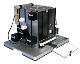
Thermal Place Preference, 2 Temperatures Choice Nociception Test (BIO-T2CT)

 Pain - Thermal Allodynia / Hyperalgesia
 Pain - Spontaneous Pain - Postural Deficit
 Pain - Mechanical Allodynia / Hyperalgesia
 Learning/Memory - Attention - Addiction
 Physiology & Respiratory Research
 Pain
Pain Metabolism
 Motor control
 Neurodegeneration
 Cross-disciplinary subjects
 Muscular system
 General activity
 Mood Disorders
 Other disorders
 Joints
 Central Nervous System (CNS)
 Sensory system
