 Douleur - Allodynie/Hyperalgésie Thermique
 Douleur - Spontanée - Déficit de Posture
 Douleur - Allodynie/Hyperalgésie Mécanique
 Apprentissage/Mémoire - Attention - Addiction
 Physiologie & Recherche Respiratoire
 Douleur
 Métabolisme
 Système moteur
 Neurodégénérescence
 Thématiques transversales
 Système musculaire
 Functions de motricité générale
 Troubles de l'humeur
 Other disorders
 Joints
 Système Nerveux Central (SNC)
  Système sensoriel
Authors
J. Davies and D. Rubinsztein.
Lab
University of Cambridge, Department of Medical Genetics, Cambridge, United Kingdom.
Journal
Human Molecular genetics
Abstract
Oculopharyngeal muscular dystrophy (OPMD) is a late-onset muscular dystrophy caused by a polyalanine expansion mutation in the coding region of the poly-(A) binding protein nuclear 1 (PABPN1) gene. In unaffected individuals, (GCG)(6) encodes the first 6 alanines in a homopolymeric stretch of 10 alanines. In most patients, this (GCG)(6) repeat is expanded to (GCG)(8-13), leading to a stretch of 12-17 alanines in mutant PABPN1, which is thought to confer a toxic gain of function. Thus, OPMD has been modelled by expressing mutant PABPN1 transgenes in the presence of endogenous copies of the gene in cells and mice. In these models, increased apoptosis is seen, but it is unclear whether this process mediates OPMD. The role of apoptosis in the pathogenesis of different muscular dystrophies is unclear. Blocking apoptosis ameliorates muscle disease in some mouse models of muscular dystrophy such as laminin _-2-deficient mice, but not in others such as dystrophin-deficient (mdx) mice. Here we demonstrate that apoptosis is not only involved in the pathology of OPMD but also is a major contributor to the muscle weakness and dysfunction in this disease. Genetically blocking apoptosis by over-expressing BCL2 ameliorates muscle weakness in our mouse model of OPMD (A17 mice). The effect of BCL2 co-expression on muscle weakness is transient, since muscle weakness is apparent in mice expressing both A17 and BCL2 transgenes at late time points. Thus, while apoptosis is a major pathway that causes muscle weakness in OPMD, other cell death pathways may also contribute to the disease when apoptosis is inhibited.
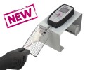
BIOSEB Instruments Used:
Grip strength test (BIO-GS3)